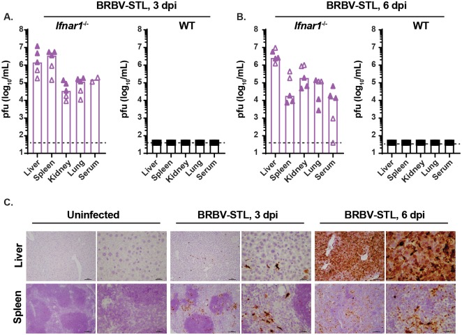
Fig 4. Liver and spleen tropism of BRBV-STL in Ifnar1-/- mice.
(A and B) Virus titers (pfu/mL) were quantified in the liver, spleen, kidney, lung and serum of WT and Ifnar1-/- mice 3 and 6 dpi after IP inoculation with 4 x 10⁴ pfu of BRBV-STL. Tissues were collected, homogenized, and the virus titer was determined by plaque assay. Each data point is a single mouse obtained from two different experiments (indicated by the open and closed symbols). The bar represents the median virus titer observed in each of the tissues. Viremia in serum at 3 dpi was obtained from two mice in one experiment. The dotted line represents the limit of detection of the assay at 40 pfu/mL. (C) RNA in situ hybridization on sections from liver and spleen from uninfected of BRBV-STL infected (4 x 10⁴ pfu of BRBV-STL via IP) Ifnar1-/- mice. Probes targeting segment 5 of BRBV were used to visualize BRBV-infected cells, indicated by the dark brown staining. Sections were counterstained with hematoxylin prior to mounting and analysis. From left to right are uninfected, 3 dpi and 6 dpi at 10x and 40x magnification. Top panel are liver sections and the bottom panel are spleen sections. The images are representative of sections obtained from three mice in one experiment.

