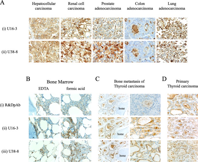
Fig 6. Novel anti-CD26 mAbs exhibit a reliable staining pattern and intensity for various tumors and decalcified specimens.
A. The tissue specimens of hepatocellular carcinoma, renal cell carcinoma, prostate adenocarcinoma, colon adenocarcinoma or lung adenocarcinoma were stained with purified novel mouse anti-human CD26 mAbs (U16-3 (i) or U38-8 (ii)). Original magnification, 40x. B, C, D. The EDTA-decalcified (left panels) or formic acid-decalcified (right panels) tissue specimens of normal human bone and bone marrow (B), formic acid-decalcified tissue specimens of metastatic thyroid carcinoma in the bone (C), or the tissue specimens of primary thyroid carcinoma without decalcification (D) were stained with purified goat anti-human CD26 pAb (R&D Systems (i)) or purified novel mouse anti-human CD26 mAbs (U16-3 (ii) or U38-8 (iii)). Original magnification, 10x (B, left panels of C, D) or 40x (right panels of C). All specimens were counterstained with hematoxylin.

