Figure 7.
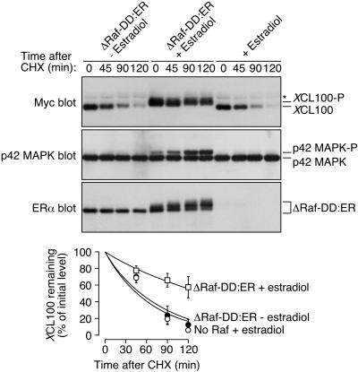
Stabilization of XCL100 follows its p42 MAPK-dependent phosphorylation in sorbitol-treated oocytes. Stage VI oocytes were microinjected with mRNA (46 ng) encoding ΔRaf-DD:ER and incubated at 16°C for 8–12 h. Oocytes were then microinjected a second time with mRNA (46 ng) encoding Myc-tagged XCL100 and were split into two pools 4 h later. Estradiol (2 μM) was added to one pool to activate Raf, and an equal volume of ethanol (0.02%) was added to the second pool as a control. Three hours later, oocytes were transferred to OR2 containing 50 μg/ml cycloheximide (CHX) plus 0.2 M sorbitol. Samples were collected at the indicated times and analyzed by immunoblotting with 9E10 (to detect XCL100), DC3 (to detect p42 MAPK), or an estradiol receptor antibody (to detect ΔRaf-DD:ER) (top three panels). Oocytes microinjected with XCL100 alone and treated with 2 μM estradiol were processed in parallel as controls to exclude the possibility of nonspecific estradiol-mediated effects. XCL100 proteins were quantified using the Kodak Image Station 440CF and 1D Image Analysis Software (bottom). Data are shown as mean values ± SEM for three independent experiments. The asterisk to the right of the Myc immunoblot denotes a nonspecific band present in samples from injected and uninjected oocytes.

