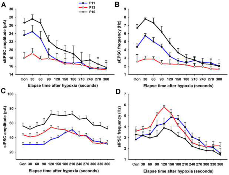
Fig. 2.

Effect of acute hypoxia on sEPSCs (A and B) and sIPSCs (C and D) of NTSVL neurons at P11 (before the critical period), P13 (during the critical period), and P15 (after the critical period). Note that the amplitude and frequency of sEPSCs were the lowest at P13 and that hypoxia induced a biphasic response at P11 and P15, but not at P13. On the other hand, the amplitude of sIPSCs at P13 was between those of the other two time-points. Hypoxia induced a rise in the frequency of sIPSCs above control levels after 1.5-3 min of exposure that was greater at P13 than at the other two time-points. (Modified from Gao et al., 2015).
