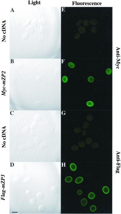
Figure 2.
Immunofluorescence staining of Myc-mZP2 and Flag-mZP3 in growing mouse oocytes. Growing oocytes were isolated from juvenile mice, as described in MATERIALS AND METHODS. After microinjection of Myc-mZP2 and Flag-mZP3 cDNA (∼1 mg/ml) into the oocyte GV, oocytes were cultured for ∼20 h, as described in MATERIALS AND METHODS. Epitope-specific monoclonal antibodies, 9E10 (anti-Myc) and M2 (anti-Flag), were used to immunolabel oocytes at the end of the culture period. Goat anti-mouse IgG coupled to FITC was used as a secondary antibody. Light images of immunolabeled oocytes are presented in A–D and fluorescent images are presented in E–H. Whereas uninjected oocytes showed no staining (A and E [anti-Myc] and C and G [anti-Flag]), injected oocytes exhibited intense fluorescence (B and F [anti-Myc] and D and H [anti-Flag]). The most intense signal was seen at the surface of the oocytes over the plasma membrane/ZP region. In each case, 100–200 oocytes were examined by LSCM. Bar (in D), 50 μm.

