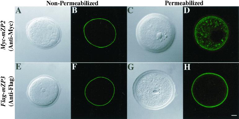
Figure 4.
Localization of Myc-mZP2 and Flag-mZP3 in growing mouse oocytes. Oocytes from 14-d-old mice were microinjected with either Myc-mZP2 or Flag-mZP3. After ∼21 h of culture, the oocytes were stained with anti-Myc (A–D) or anti-Flag (E–H), followed by FITC-conjugated secondary antibody. Light (A, C, E, and G) and fluorescent (B, D, F, and H) images of oocytes are presented. In fixed, permeabilized oocytes, both epitope-tagged ZP proteins were detected, primarily over the plasma membrane/ZP region (D and H). In addition, intracellular vesicles were readily observed in Myc-mZP2-injected oocytes (D). Much less intracellular staining was detected in Flag-mZP3-injected oocytes (H), although in some cases, vesicles were detected. The plasma membrane/ZP staining appears to be on the outside of permeabilized growing oocytes, because fixed, nonpermeabilized samples (A, B, E, and F) exhibited very similar staining patterns. In each case, 100–200 oocytes were examined by LSCM. Bar (H), 10 μm.

