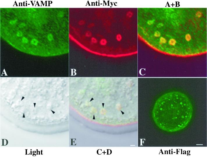
Figure 5.
Colocalization of Myc-mZP2 and VAMP in growing mouse oocytes. Oocytes from 14-d-old mice were microinjected with Myc-mZP2. After ∼21 h of culture, the oocytes were stained with anti-VAMP (IgM; A) and anti-Myc (IgG; B), followed by FITC-conjugated rabbit anti-mouse IgM and Texas Red-conjugated goat anti-mouse IgG secondary antibody. Light (D) and fluorescent (A–C) images of oocytes are presented. C, An overlap of A and B. Note the presence of VAMP (green) and Myc-mZP2 (red) in the large, doughnut-shaped vesicles present in the cortical region of the oocyte; overlap of VAMP and Myc-mZP2 is yellow (green plus red). E, An overlap of C and D. Arrowheads in D and E, the positions of large vesicles in the cortical region of the oocyte. In F, an oocyte microinjected with mZP2 tagged with Flag at its C terminus (Myc-mZP2-Flag) and probed with anti-Flag is shown. Note that the vesicles exhibit fluorescence along their membranes. In each case, 100–200 oocytes were examined by LSCM. Bars (E and F), 2 μm and 10 μm, respectively.

