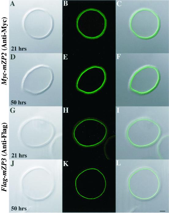
Figure 6.
Assembly of Myc-mZP2 and Flag-mZP3 into ZP of growing mouse oocytes. Either Myc-mZP2 (A–F) or Flag-mZP3 (G–L) cDNA was microinjected into growing oocytes isolated from 13-d-old mice. After the oocytes were cultured for ∼21 (A–C and G–I) and ∼50 h (D–F and J–L), ZP were isolated in the presence of 1% NP-40 and immunolabeled with either anti-Myc or anti-Flag and FITC-conjugated secondary antibody. Light images are shown in A, D, G, and J and fluorescent images are shown in B, E, H, and K. Composites of light and fluorescent images are shown in C, F, I, and L. In all cases, epitope-tagged ZP glycoproteins were found only at the innermost layer of the ZP (A–F, Myc-mZP2; G–L, Flag-mZP3). The intensity of the immunofluorescence signal increased with increasing culture times (compare B and E, Myc-mZP2; compare H and K, Flag-mZP3; see text for details). More than 200 isolated ZP were stained in this manner. Bar (L), 10 μm.

