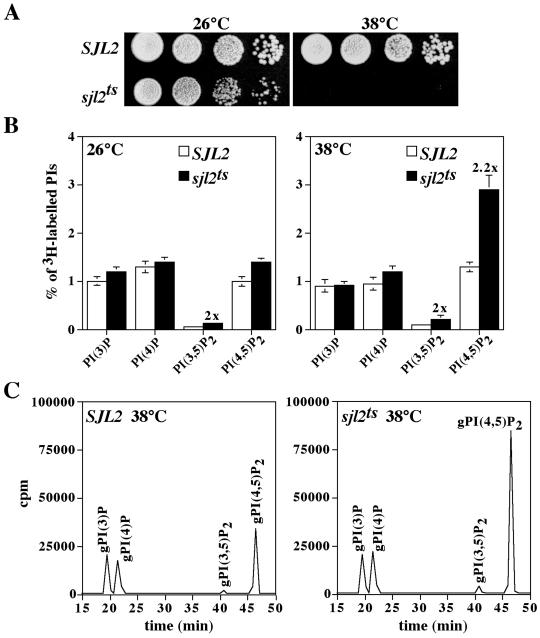
Figure 2.
sjl1Δ sjl2ts sjl3Δ cells display growth defects and generate increased levels of PtdIns(4,5)P2 at the restrictive temperature. (A) sjl1Δ sjl2Δ sjl3Δ cells expressing either SJL2 (YCS157) or sjl2ts (YCS176) from LEU2 CEN plasmids were grown in YPD at the permissive temperature. Tenfold serial dilutions of cells were spotted onto YPD and grown for 2 d at 26°C or 38°C. (B and C) Cells were preincubated at either 26°C or 38°C for 10 min and labeled with myo-[2-3H]inositol for 50 min at the permissive or nonpermissive temperatures. Lipids were then deacylated from cellular membranes, and glycero-phosphoinositols were extracted and analyzed by HPLC. (B) Quantitative comparisons of glycero-phosphoinositols generated by sjl1Δ SJL2 sjl3Δ cells (YCS157; open columns) and sjl1Δ sjl2ts sjl3Δ cells (YCS176; black columns) at 26°C (left) and 38°C (right) are shown. These data represent the means ± SD of three independent experiments. (C) Typical HPLC elution profiles are shown for sjl1Δ SJL2 sjl3Δ cells (YCS157; left) and sjl1Δ sjl2ts sjl3Δ cells (YCS176; right) at 38°C. Peaks corresponding to glycero-Ins(3)P, glycero-Ins(4)P, glycero-Ins(3,5)P2, and glycero-Ins(4,5)P2 are indicated.

