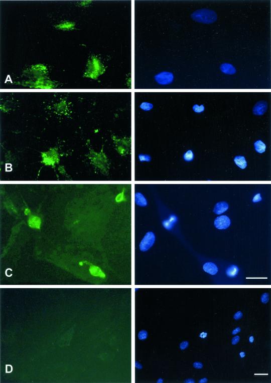
Figure 3.
Macromolecular internalization after drug treatment or low temperature. Right column shows Hoechst staining; left column shows FITC staining. REF52 cells were treated either for 1 min with 0.25% trypsin, for 30 min with 10 μM cytochalasin D, or for 30 min with 5 μM nocodazole, before application of FITC-dextrans for 15 min at 37°C, rinsed, and fixed as described in Figure 1. For cytochalasin D or nocodazole treatments, the drug was also included at the same concentration together with the dextrans during the 15-min incubation time. Alternatively, cells were placed at 4°C for 30 min and further incubated at 4°C in the presence of FITC-dextran for 60 min. (A) Fluorescence of 20-kDa dextran after incubation onto trypsinized cells. (B) Fluorescence of 20-kDa FITC-dextran after incubation onto cytochalasin D-treated cells. (C) Fluorescence of 40-kDa FITC-dextran after incubation onto nocodazole-treated cells. (D) Fluorescence uptake of 20-kDa FITC-dextran after incubation at 4°C for 60 min. Bar, 10 μm.

