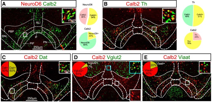
Figure 4.
NeuroD6 mRNA co-localizes partly with Calb2 mRNA, but Calb2 mRNA is abundant throughout the VTA and SNc. Double-labeling FISH in the ventral midbrain of adult wild-type mice detecting the following mRNAs. A, NeuroD6 (red) and Calb2 (green), inset with high magnification of overlap (yellow), pie charts illustrating quantification of overlap between NeuroD6 and Calb2. B, Calb (red) and Th (green), inset with high magnification of overlap (yellow), pie charts illustrating quantification of overlap between Th and Calb2. C, Calb2 (red) and Dat (green), inset with high magnification of Dat/Calb2 mRNA overlap (yellow), pie chart illustrating quantification of overlap between Th and Calb2. D, Calb2 (red) and Vglut2 (green), inset with high magnification of Vglut2/Calb2 mRNA overlap (yellow) in blue square and Vglut2-negative/Calb2-positive (red) in white square, pie chart illustrating quantification of overlap between Th and Calb2. E, Calb2 (red) and Viaat (green), inset with high magnification of Viaat negative or positive (red, yellow, green) in white square. VTA, ventral tegmental area; SNc, substantia nigra pars compacta; PBP, parabrachial pigmented nucleus; PN, paranigral nucleus; PIF, parainterfascicular nucleus; RLi, rostral linear nucleus; IF, interfascicular nucleus. Calb2, Calbindin 2 (Calretinin); Dat, Dopamine transporter; Th, Tyrosine hydroxylase; Vglut2, Vesicular glutamate transporter 2; Viaat, Vesicular inhibitory amino acid transporter; FISH, fluorescent in situ hybridization.

