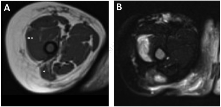
FIG E1. Magnetic resonance imaging (MRI) of upper thighs showing muscular atrophy and patchy myositis.
(A) T1 weighted axial MRI of right and left thigh shows atrophy and fatty replacement of the gluteus maximus and vastus medialis muscles. (B) The corresponding T2 weighted fat suppressed axial image shows patchy enhancement in these areas suggestive of active inflammation.

