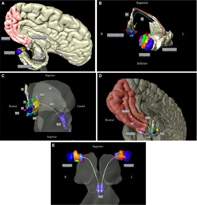
Figure 1.
Schematic representation of the participation of the amygdala and hypothalamus in the neurocircuitry underlying aggressive behavior. Overview of A, the main structures implicated in the control of aggressive behavior and B, the main connections between the hypothalamus and amygdala; C, between the hypothalamus and PAG; D, among the amygdala, hypothalamus, and frontal cortex; and E, between the amygdala and PAG. The 3-dimensional reconstructions are based on histological segmentations of the depicted structures (methods described in Alho et al144). OMPFC: orbitomedial prefrontal cortex; PAG: periaqueductal gray; Fx: fornix; St: stria terminalis; Hyp: hypothalamus; So: supraoptic nucleus; Pv: paraventricular hypothalamic nucleus; MB: mammillary body; Mmt: mammillothalamic tract; Th: thalamus; DLF: dorsal longitudinal fasciculus; MFB: medial forebrain bundle; UF: uncinate fasciculus.

