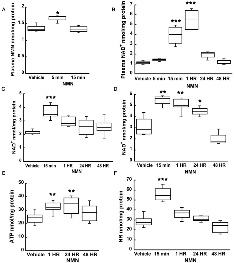
Figure 3.
NMN administration leads to increase in plasma and hippocampal tissue NAD+, NMN, NR, and ATP levels. (A) Plasma NMN levels increased at 5 min after NMN injection and normalized to vehicle by 15 min. (B) There was a significant increase in plasma NAD+ levels, at 15 min and at 1 hour after NMN treatment. Twenty-four hours following NMN injection the NAD+ levels returned to vehicle. (C) Hippocampal tissue NAD+significantly increased also already at 15 min after NMN administration but afterwards begin to decline back to physiological levels. (D) In isolated hippocampal mitochondrial, NAD+ increased by 76% from 15 min to 24 hours following NMN injection. (E) Hippocampal tissue showed an increase in ATP levels at 1 hour that was sustained up to 24 hours after NMN administration. (F) Levels of NAD+ precursor, NR, increased by 60% at 15 min NMN post-injection in hippocampal tissue suggesting that at least part of NMN is metabolized into NR. * p<0.05, ** p<0.01, *** p<0.001 compared to vehicle (n=11 (A), n=26 (B), n=30 (C), n=20 (D), n=22 (E), n=25 (F)) (One-way ANOVA followed by Tukey HSD test).

