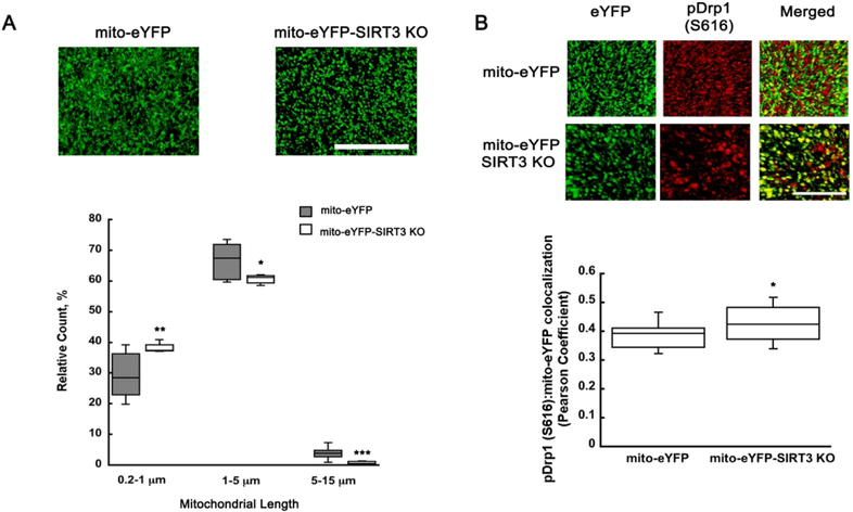
Figure 7.
pDrp1 (S616) shows increased colocalization with neuronal mitochondria leading to more fragmented organelles in SIRT3KO mice. Z-stack images from hippocampal CA1 oriens of mito-eYFP and mito-eYFP-SIRT3KO brain sections were collected and analyzed using Volocity software. (A) The fragmented mitochondrial population (0.2-1 μm) increased by 27% when compared to vehicle treated animals, while the rod shape (1-5 μm) and tubular (5-15 μm) population decreased 8% and 74%, respectively. Mito-eYFP and mito-eYFP-SIRT3KO brain sections were stained with pDrp1 (S616) (red) and colocalization between pDrp1 (S616) and eYFP analyzed using Volocity software. SIRT3KO samples show an increase in colocalization of pDrp1 (S616) and mito-eYFP (green) resulting in increased fragmentation (B). Scale bars represent 100 μm. *p<0.05, **p<0.01, ***p<0.001 compared to mito-eYFP (n=27 (A), n=31 (B)) (A Student T-test, B One-way ANOVA followed by Tukey HSD test)

