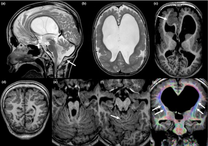
Figure 1.

Sagittal turbo spin‐echo T2‐weighted sequence (a) showing macrocrania, dysmorphic skull base, anti‐clockwise rotation and hypoplasia of the vermis, brainstem hypoplasia, and stretched corpus callosum; a dysmorphic skull base with thickened clivus, crista galli and posterior margin of the foramen magnum (asterix and arrow, respectively) is also evident. Bilateral polymicrogyria and ventriculomegaly are shown on axial turbo spin‐echo T2‐weighted image (b). Axial reformation of 3D turbo field echo T1 weighted sequence showing dysmorphic basal ganglia and pronounced infolding and thickening of the right frontal cortex (arrow) (c). Coronal reformation of 3D turbo field echo T1 weighted sequence showing parieto‐occipital polymicrogyria (d). Dysmorphic midbrain and disorganized foliation [arrow on (f)] on axial reformation of 3D turbo field echo T1 weighted sequence (e and f). Coronal reformats of colored Fractional Anisotropy map from Diffusion Tensor Imaging showing white matter hypoplasia and fascicle abnormalities, including laterally displaced cortico‐spinal tracts (arrows) and absence of superior longitudinal fasciculus (g)
