Figure 2.

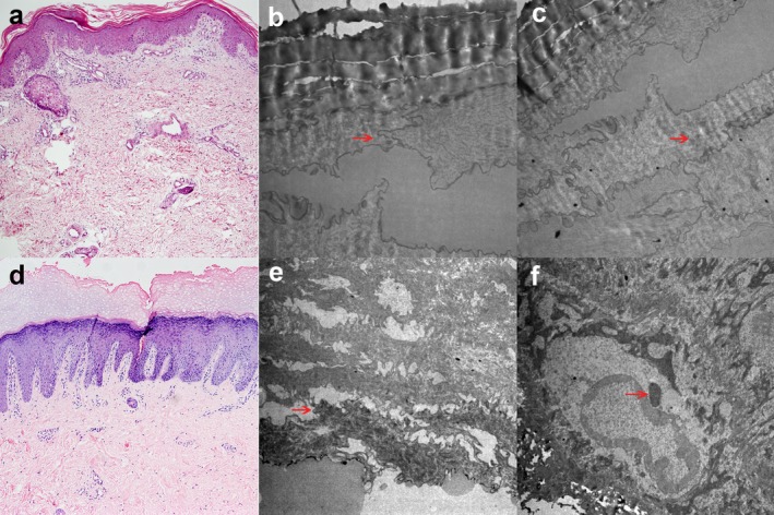
Histologic features of EKVP. (a) Histology of affected skin in patient 1 revealed papillomatosis, acanthosis, hypergranulosis, compact orthohyperkeratosis, parakeratosis, and follicular plugging (×100, H&E stain). (b) The electron microscope (EM) showed stratum corneum thickening, filaments tearing (red arrowheads) (×10,000), (c) and filaments of interstellar clouds (red arrowheads) (×8,000). (d) Histology of affected skin in patient 2 revealed compact orthohyperkeratosis, vacuolated granular cells with sparse keratohyalin granules, acanthosis, papillomatosis and perivascular infiltrates (×100, H&E stain). (e) The EM showed stratum corneum thickening, filaments tearing (red arrowhead) (×8,000), (f) the cytoplasm and nucleus edema of hyaline layer cells, superficial granular cells containing characteristic intranuclear granules (red arrowhead) (×10,000)
