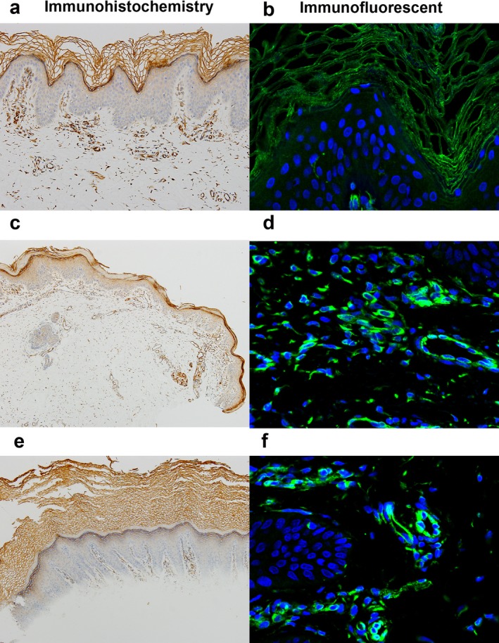
Figure 4.

Immunohistochemistry and immunofluorescent staining of Cx43. (a, b) In control healthy skin, immunosignals of anti‐Cx43 were expressed in the intracellular junctions of keratinocytes in the stratum corneum and granular layer (×100, DAB stain, ×200). (c) In lesion of patient 1, immunosignals of anti‐Cx43 were strongly expressed in the stratum corneum, granular layer and spinous layer, whereas basal layer show low expression of Cx43 (×100, DAB stain). (d) Cx43 protein was mainly expressed in the cytomembrane and cytoplasm (×200). (e) In lesion of patient 2, immunosignals of anti‐Cx43 were strongly expressed in the stratum corneum and granular layer (×100, DAB stain). (f) Cx43 protein was mainly expressed in the cytomembrane and cytoplasm (×200)
