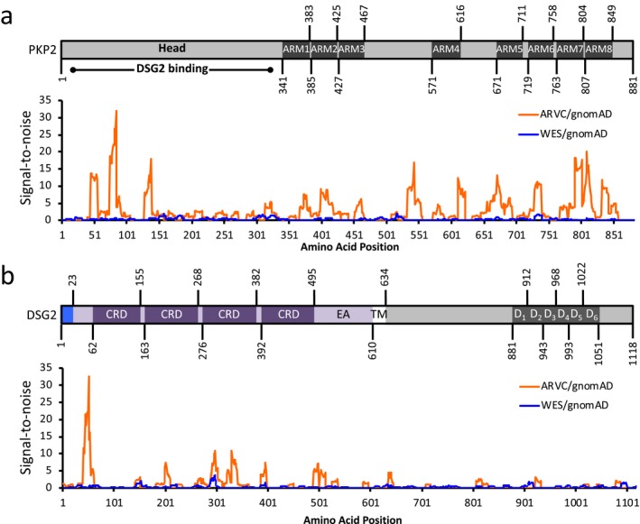
Figure 5.

Topological signal‐to‐noise (S:N) mapping between WES (blue line) and ARVC (orange line) cohorts, as normalized against gnomAD for (a) PKP2 (NP_001005242.2), and (b) DSG2 (NP_001934.2). Accompanying protein topologies are shown to scale. Intracellular regions (grey fill), extracellular regions (purple fill), transmembrane regions (white fill), and signal pro‐peptides (light blue fill), are represented. ARM, armadillo/beta‐catenin‐like repeat. ARVC, arrhythmogenic right ventricular cardiomyopathy. CRD, Cadherin repeat domain. D1–6, DSG repeat domains 1–6. EA, extracellular anchor. Head, head domain. TM, transmembrane domain. WES, whole exome sequencing
