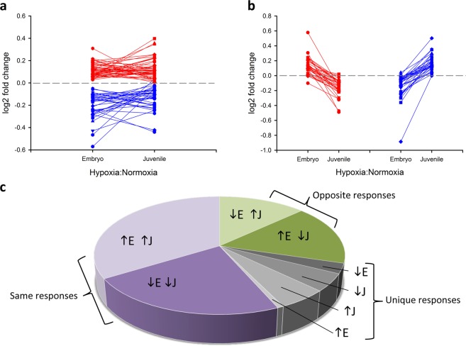
Figure 1.
Patterns of protein expression changes induced by developmental hypoxic exposure in embryonic (E, 90% incubation) and juvenile (J, 2 y old) alligator hearts. (a) Differentially abundant proteins with p < 0.05 for the main effect of oxygen, where red are up-regulated and blue are down-regulated proteins, expressed as log2 fold change from the normoxic controls. (b) Differentially abundant proteins with p < 0.05 for the interaction effect of oxygen × age, where red traces are proteins with higher expression in embryonic than juvenile hearts, and blue traces indicate the opposite trend, in log2 fold change from normoxic controls. (c) Proportional representation of the 145 differentially abundant proteins, grouped as those with similar direction of response in embryonic and juvenile hearts (purple sections), those with opposite direction of response between the two ages (green sections), and those proteins that were uniquely regulated in a single treatment (grey sections).

