Fig. 1.
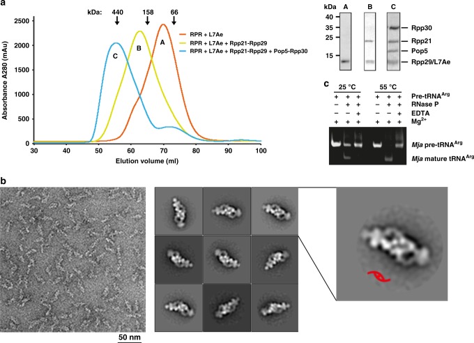
In vitro reconstitution of the MjaRNase P holoenzyme. a Size exclusion chromatographic profiles (left) and Coomassie-stained SDS-PAGE of the corresponding peaks in the profiles (right). b Negative staining EM analysis of MjaRNase P. Left: A representative negative-stained EM micrograph of MjaRNase P. Middle: Selected 2D class averages of MjaRNase P. Right: Close-up view of the 2D class averages with the C2 symmetry denoted a red symbol. c In vitro pre-tRNAArg processing assay of the MjaRNase P holoenzyme

