Fig. 2.
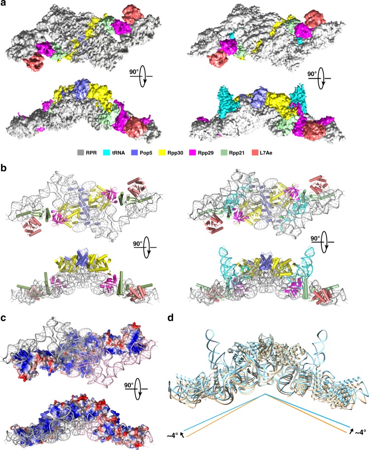
Overall structures of the MjaRNase P complex with or without tRNA. a The cryo-EM density maps of MjaRNase P (left) and the MjaRNase P-tRNATyr complex (right) are shown in two orthogonal views. Protein and RNA components are color-coded and the scheme is shown below the figure. b Two orthogonal views of the atomic model of MjaRNase P (left) and the MjaRNase P-tRNA complex (right) are shown in cartoon representation. Protein and RNA components are color-coded as in (a). c Two orthogonal views of the surface electrostatic potential of the protein assembly in MjaRNase P reveals a continuous highly basic surface that binds two RPRs (negative: red; positive: blue). The two RPRs are colored in pink and gray, respectively. d Superposition of the structures of MjaRNase P with or without tRNA shows that tRNA binding only induces a ~8° change in the angle between the two MjaRNase P monomers. MjaRNase P and the MjaRNase P-tRNA complex are colored in wheat and palecyan, respectively

