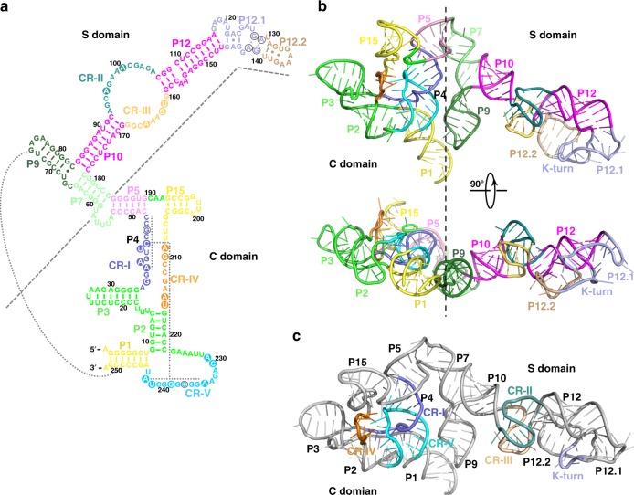
Fig. 3.
Structure of the M. jannaschii RPR. a Secondary structure of RPR. Structural elements are colored to match their labels. The conserved regions of RPR (CR-I to CR-V) are colored in slate, deepteal, yelloworange, orange and cyan, respectively. The conserved P4 stem is denoted with dotted line. Dotted line between stem P1 and P9 indicates long-range RNA-RNA interaction. b Two orthogonal views of the overall structure of RPR. RPR is colored as in (a). c Overall structure of the RPR. The pseudoknot and the T-loop regions locate in the C and S domains of RPR, respectively. CR-I to CR-V are colored as in (a)

