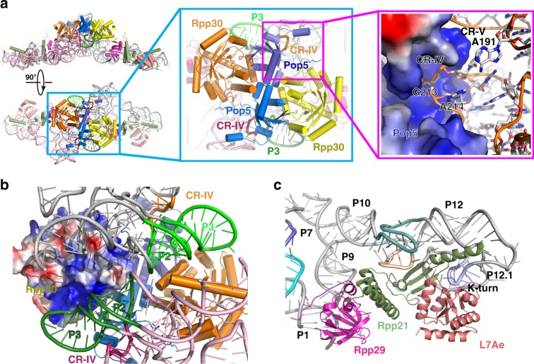
Fig. 5.
Interaction between RPR and RPPs in MjaRNase P. a Left: The atomic model of the MjaRNase P complex is shown in two orthogonal views. RPPs are color-coded as in Fig. 2a. The two RPR molecules are colored in pink and gray, respectively. Middle: Close-up view of the interface between two RPRs (colored in pink and gray) and the (Pop5-Rpp30)2 heterotetramer. The two P3 stems are colored in deepteal and green, respectively. Right: Close-up view of the interface between Pop5 and CR-IV of RPR. Pop5 is shown in electrostatic surface representation and RPR in cartoon representation. b Rpp30 simultaneously interacts with the two RPR molecules in the dimeric holoenzyme. One Rpp30 is shown in electrostatic surface representation and the interacting P2-P3 stem from one RPR is colored in deepteal and the other interacting RPR in green. c Close-up view of the interface between the Rpp29-Rpp21-L7Ae heterotrimer and RPR. RPR and RPPs are shown in cartoon and colored as in Fig. 3c and Fig. 2a, respectively

