Fig. 6.
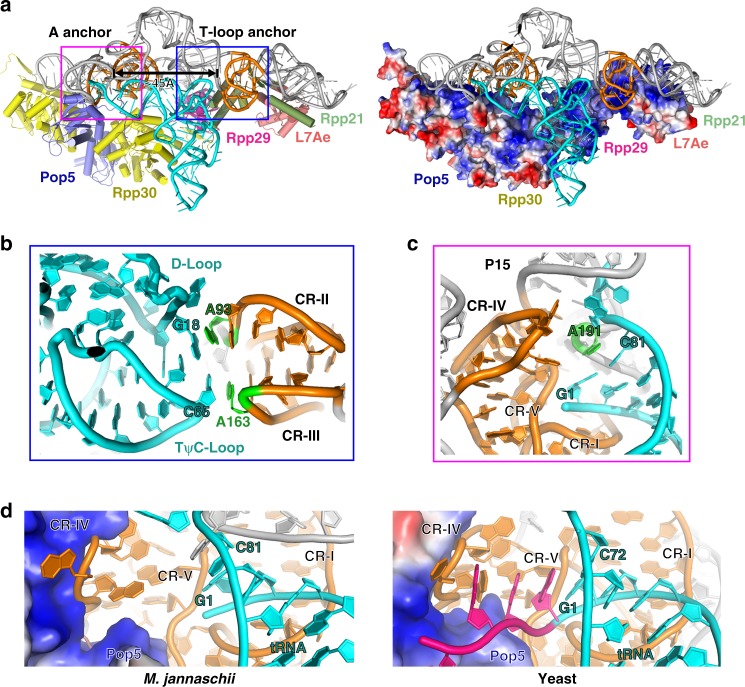
tRNA recognition by MjaRNase P. a Overall structure of the monomeric MjaRNase P complex bound with tRNA. Protein components are shown in cartoon (left) and in electrostatic surface (right) representations, respectively. The magenta box denotes the A anchor and the blue box denotes the T-loop anchor. The distance between the two anchors is highlighted with a black double arrow. The tRNA is colored in cyan. b Close-up view of the TψC and D loops of tRNA that stack with the T-loop anchor in CR-II and CR-III of RPR. c Close-up view of the central nucleotide A191 (in green) in loop L5–15 of RPR that stacks on the first base-pair G1-C81 of tRNA. d Left panel: Close-up view of the active site in the MjaRNase P-tRNA complex. Right panel: Close-up views of the active site in the yeast RNase P-pre-tRNA complex (PDB: 6AH3). Pop5 is shown in surface representation and colored in electrostatic potential. RPR and tRNA are shown in cartoon and colored in orange and cyan, respectively. The 5′ leader of yeast pre-tRNAPhe is colored in magenta

