Fig. 8.
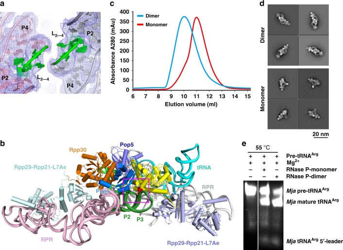
Dimeric organization of MjaRNase P. a The loops between stems P2 and P4 from the two RPRs staggered pack together. The two loops are colored in green and the two RPRs in pink and gray, respectively. The EM density map is shown in light blue mesh. b Overall view of the dimeric conformation mediated by the (Pop5-Rpp30)2 heterotetramer. Rpp30 simultaneously interacts with the two RPR molecules in the dimeric holoenzyme. c SEC profiles of the WT (Dimer, blue) and mutant (Monomer, red) MjaRNase P complexes. d Selected 2D class averages of the WT (Dimer, top) and mutant (Monomer, bottom) MjaRNase P complexes. e In vitro tRNA processing assay of WT (Dimer) and mutant (Monomer) MjaRNase P complexes

