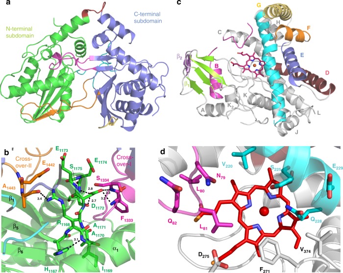
Fig. 6.
Structure of individual domains within the OxyAkis/Xkis complex. a Structure of the X-domain from kistamicin biosynthesis (coloured as in Fig. 5). b Typical active site region of a C/E-type domain showing the effects of mutating the conserved HHxxxDG motif into the HRxxxDE motif found in Xkis (coloured as in Fig. 5, side chains shown as sticks). c Structure of the OxyAkis enzyme (B-helix and loop shown in magenta, D-helix shown in firebrick red, E-helix shown in blue, F-helix shown in orange, G-helix shown in yellow, I-helix shown in cyan, β-1 region shown in green, β-2 region shown in purple, heme shown in red sticks). d Active site of OxyAkis (coloured as in c, side chains shown as sticks)

