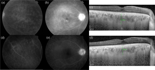
Figure 3.
Fluorescein angiography (FA) and enhanced depth imaging optical coherence tomography (EDI-OCT) images of the active and convalescent phases in a representative eye with Behçet’s disease. In the active phase, FA reveals dense retinal vascular leakage in all retinal locations (peripheral retina (a), macula (b), and optic disc (b)), and the subfoveal choroidal thickness (SCT) was determined to be 453 μm (c). In the convalescent phase, the retinal vascular leakage improved in all parts (d,e), while the SCT was decreased at 364 μm (f).

