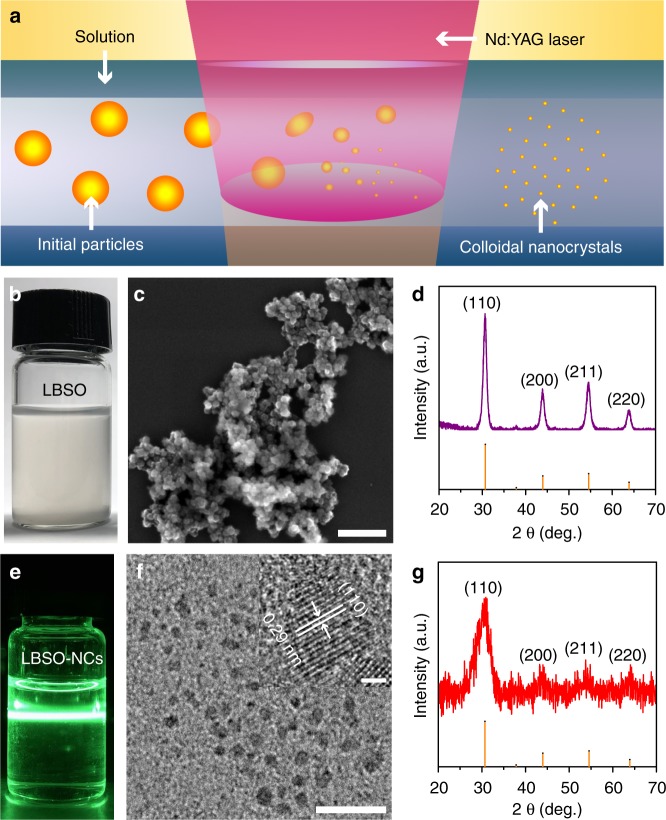
Fig. 1.
LBSO before and after LSPC. a Schematic illustration of LSPC. b Optical image of LBSO particles dispersed in the solvent before LSPC. c SEM image and d XRD pattern of LBSO particles fabricated. e Mie-scattering image of laser generated LBSO colloidal nanocrystals (LBSO-NCs). f TEM and HRTEM image (insert) of LBSO nanocrystals generated by pulsed laser irradiation. g XRD pattern of LBSO nanocrystals after pulsed laser irradiation. Scale bars: (c) 250 nm, (f) 20 nm and insert of (f) 2 nm

