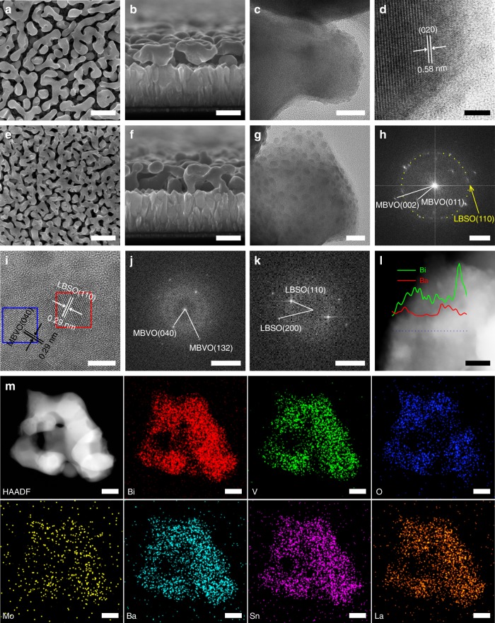
Fig. 2.
Characterizations of MBVO and LBSO-MBVO films. a SEM top-view, b SEM cross-section, c TEM, and d HRTEM image of MBVO films. e SEM top-view, f SEM cross-section, g TEM image, h FFT-transformation image and i HRTEM image of LBSO-MBVO-2 film. j FFT-transformation image of MBVO matrix (area labeled in blue in Figure i). k FFT-transformation image of LBSO nanocrystal (area labeled in red in Figure i). l HAADF image and corresponding EDX-line scanning of LBSO-MBVO-2 film. m TEM-EDS analysis of LBSO-MBVO-2 film. Scale bars: (a, e) 500 nm, (b, f) 250 nm, (c, g) 20 nm, (d, i) 5 nm, (h) 2 1/nm, (j, k) 5 1/nm, (l) 10 nm and (m) 100 nm

