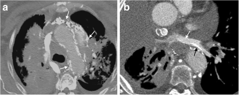
Fig. 12.

Unusual collaterals. a Axial CT image of a patient with numerous unnamed collateral vessels coursing through the mediastinum (white arrows). b Axial CT scan demonstrates collateral vessels (black arrow) draining into the left pulmonary veins (white arrow)
