Fig. 4.

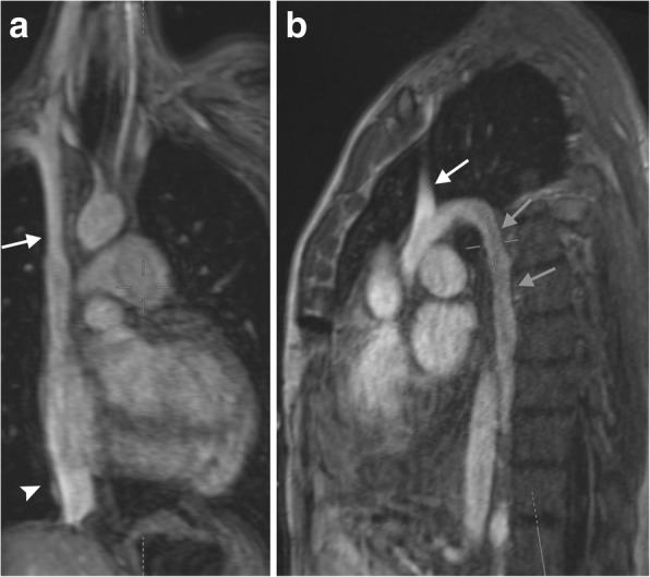
MRI venous imaging. Coronal (a) and sagittal (b) images of the thoracic venous anatomy in the late arteriovenous phase of imaging of a MRI scan. The SVC (white arrow), IVC (arrowhead), and azygos vein (gray arrows) are labeled

MRI venous imaging. Coronal (a) and sagittal (b) images of the thoracic venous anatomy in the late arteriovenous phase of imaging of a MRI scan. The SVC (white arrow), IVC (arrowhead), and azygos vein (gray arrows) are labeled