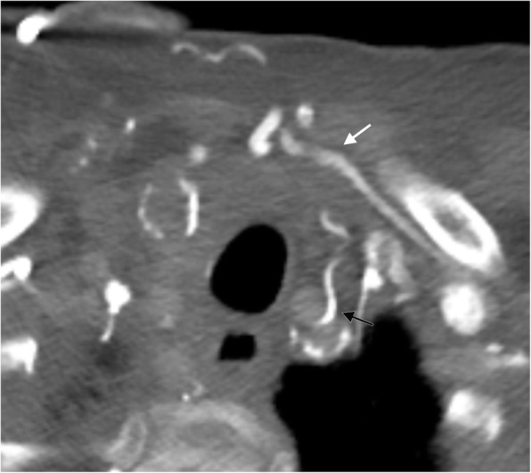
Fig. 7.

Anterior neck collaterals. Axial CT image showing the jugular venous arch (white arrow) assisting in the diversion of blood flow to the patent contralateral side and an enlarged inferior thyroid vein collateral (black arrow) in a patient with an obstructed internal jugular vein
