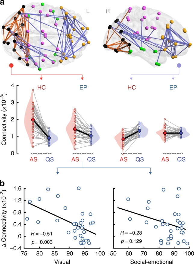
Fig. 4.

Alpha-band cortical networks showing a sleep-by-group interaction and their association with neurocognitive outcome at two years. a A positive interaction (changes in connectivity between AS and QS in HC vs. EP) is found for patterns of connectivity linking occipital regions, with only a few connections to temporal and central cortices (orange; pFWER = 0.003, paired two-tailed t-test). This pattern is spatially consistent also across neighboring theta and low beta frequency bands (Supplementary Fig. 2a). A negative interaction (opposite changes in connectivity between AS and QS as a function of group) is observed for long-range connectivity between the frontal cortex and occipital regions (violet; pFWER = 0.0004, paired two-tailed t-test). These interactions reflect attenuated changes in connectivity between sleep states in EP compared to HC. Gray lines link individual infants and thick black lines indicate slopes between group means. b Correlations between changes in the mean cortical connectivity strength of the occipital pattern (connectivity strength of the orange network of panel a in AS minus QS) and neurodevelopmental outcome assessed at two years of age in EP infants. Changes in alpha connectivity correlate significantly with visual performance (R = −0.514, p = 0.003, surviving FDR, partial two-tailed Pearson’s correlation). The association with social-emotional outcome is not statistically significant. Source data are provided as a Source Data file
