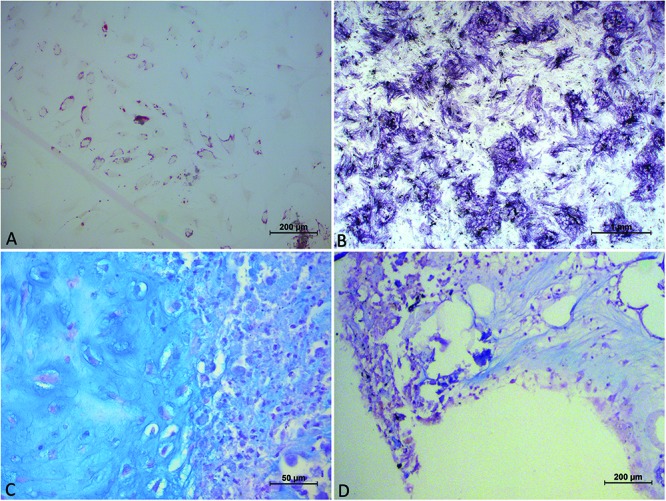
FIGURE 4.

Image showing panel (A) cells, obtained from collagenase digestion, in adipogenic medium, exhibiting the morphology of multi-vacuolar adipocytes (arrows; Scale bar: 200 μm); (B) cells, obtained from collagenase digestion, in osteogenic medium. Formation of mineralized matrix was detected by Von Kossa staining (arrows; Scale bar: 1 mm); (C) cells, obtained from collagenase digestion, in chondrogenic medium demonstrated by Alcian Blu staining; (D) cells, obtained from Rigenera digestion, in chondrogenic medium demonstrated by Alcian Blu staining.
