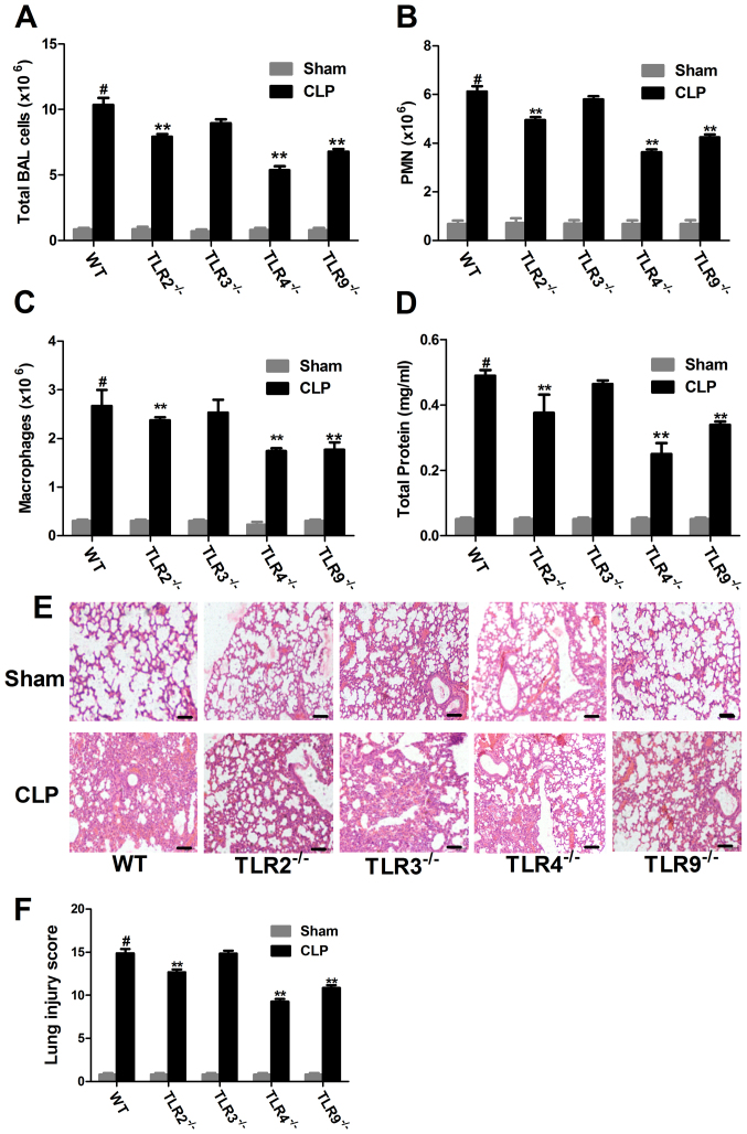
Figure 3.
Immune cell infiltration and lung injury in CLP-induced ALI in WT and TLR−/− mice. BALF samples were collected from WT and TLR−/− mice 20-h post-surgery and the (A) total, (B) PMN and (C) macrophage count was examined in each group. (D) BALF protein concentration was examined. (E) Morphological changes were observed following H&E staining in lung tissue sections (magnification, ×200). Scale bar, 200 µm. Data from at least three independent experiments. (F) CLP-induced lung injury scores were examined. Data are presented as the mean ± standard error of the mean (n=5). #P<0.05 vs. the WT sham group; **P<0.01 vs. WT CLP group. CLP, cecal ligation and puncture; ALI, acute lung injury; WT, wild-type; TLR, Toll-like receptor; PMN, polymorphonuclear cells; BALF, bronchoalveolar lavage fluid; H&E, hematoxylin and eosin.

