Figure 5.
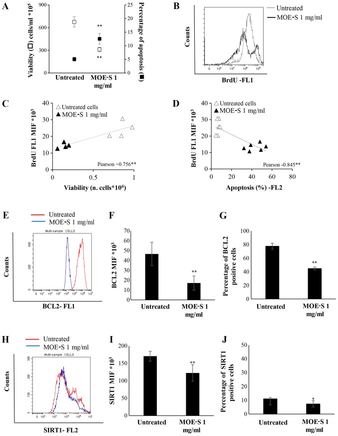
Characterization of the pro-apoptotic and anti-proliferative effects of MOE-S. (A) Jurkat cells were treated with 1 mg/ml FW of MOE boiled seeds for 72 h. Viability and apoptosis were analysed and reported as number of alive cells ×105/ml (white square) and percentage of apoptosis (black square) respectively. The reduction of DNA synthesis was characterized by bromodeoxyuridine assay: One representative overlay histogram (B) of BrdU-positive cells in untreated cells (grey line) and MOE-S (black line), analysed by Flow cytometry. Correlation analysis of DNA synthesis with respect to (C) cell viability and (D) apoptosis. (E) Representative overlay histogram of BCL2 protein expression. (F) Median intensity fluorescence (MIF) of BCL2 positive cells from three independent biological experiments. (G) The percentage of BCL2 positive cells was represented by a histogram of three independent biological experiments. (H) BCL2 overlay histogram of SIRT1 protein expression, (I) MIF of SIRT1 positive cells and (J) percentage of SIRT1 positive cells. Data are reported as the mean ± SD of three independent experiments performed. *P<0.05, **P<0.01, treated vs. untreated cells.

