Figure 1.
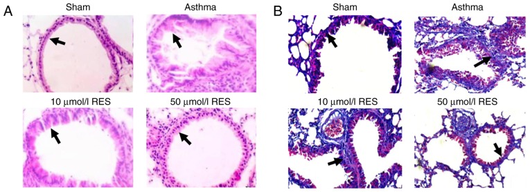
Hematoxylin and eosin, and 2 of rat lung tissues demonstrating pathological lesions and collagen deposition following establishment of an asthma rat model. (A) Pathological lesions in rat lung tissue of sham, asthma, 10 µmol/l RES and 50 µmol/l RES groups. (B) Alterations of airway collage deposition (blue staining) in rat lung tissues of sham, asthma, 10 µmol/l RES and 50 µmol/l RES groups (magnification, ×200). RES, resveratrol.

