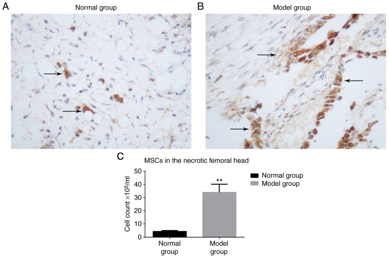
Figure 3.
Immunohistochemistry of MSCs. Immunohistochemistry staining of the (A) normal and (B) model groups (arrows indicate 5-bromo-2-deoxyuridine-labeled MSCs in the femoral head; magnification, ×200). (C) The number of MSCs present were counted and the results are indicated as a bar graph. **P<0.05 vs. normal group. MSC, mesenchymal stem cell.

