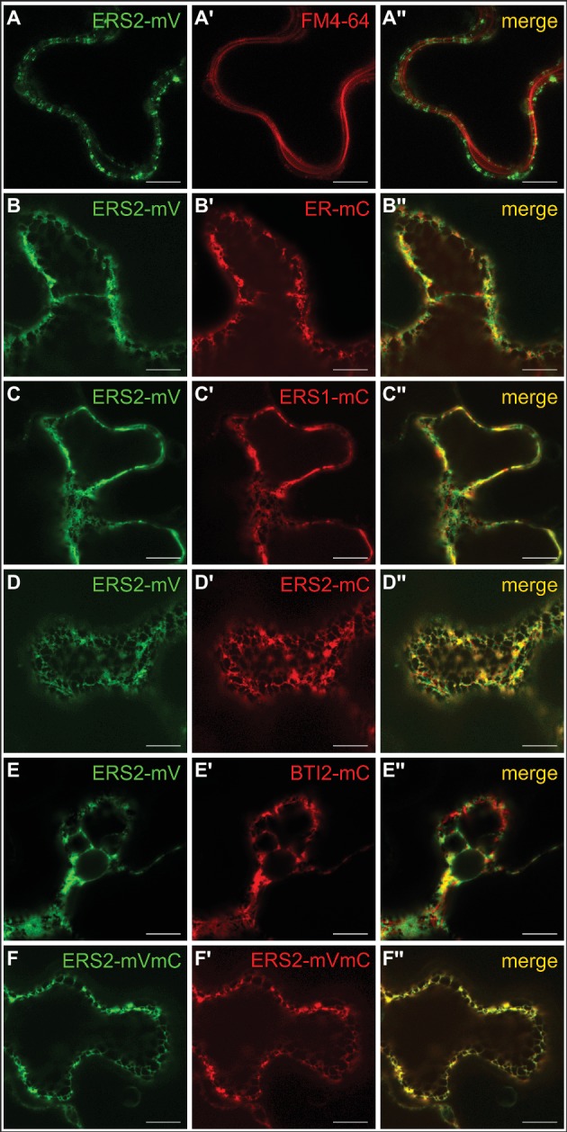
Figure 7.

Intracellular localization of AtERS1 and AtERS2 transiently expressed in N. benthamiana epidermis cells. Confocal laser scanning microscopy images of mVenus (mV) and mCherry (mC)-tagged receptor proteins. (A–A″) AtERS2 does not colocalize with the PM dye FM4-64 and is instead found (B–B″) at the ER, where colocalization with the ER-mCherry marker protein is detected. AtERS2-mVenus colocalizes with (C–C″) AtERS1-mCherry, (D–D″) AtERS2-mCherry and (E–E″) BTI2-mCherry at the ER. (F–F″) AtERS2 tagged to mVenus and mCherry is also detected at the ER. Bars = 10 μm.
