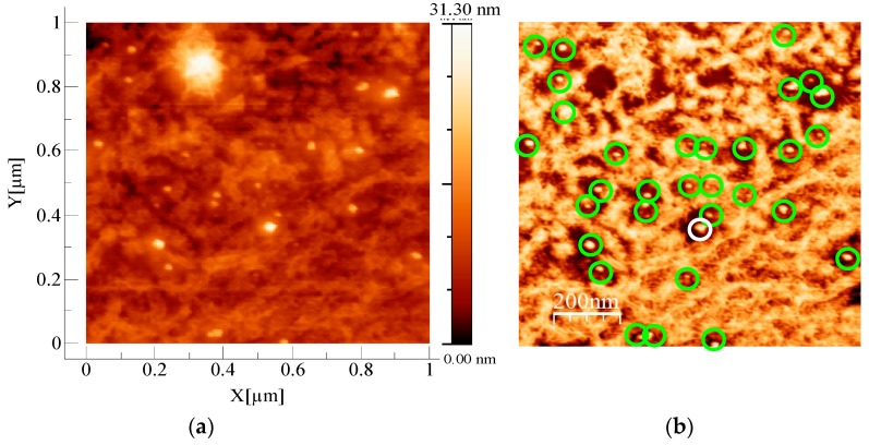
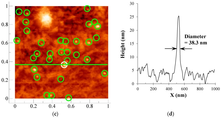
Figure 2.
(a) Atomic force microscopy (AFM) image of hybrid cellulose film (1000 nm × 1000 nm) in topographic mode; (b) AFM image of hybrid cellulose film, the same as in (a) in phase mode; circles were drawn around the undoubtedly silver nanoparticles; (c) AFM image of (a) superposed with the same circles which were drawn in the phase image in (b); (d) Height profile for a horizontal line crossing the white circle center.


