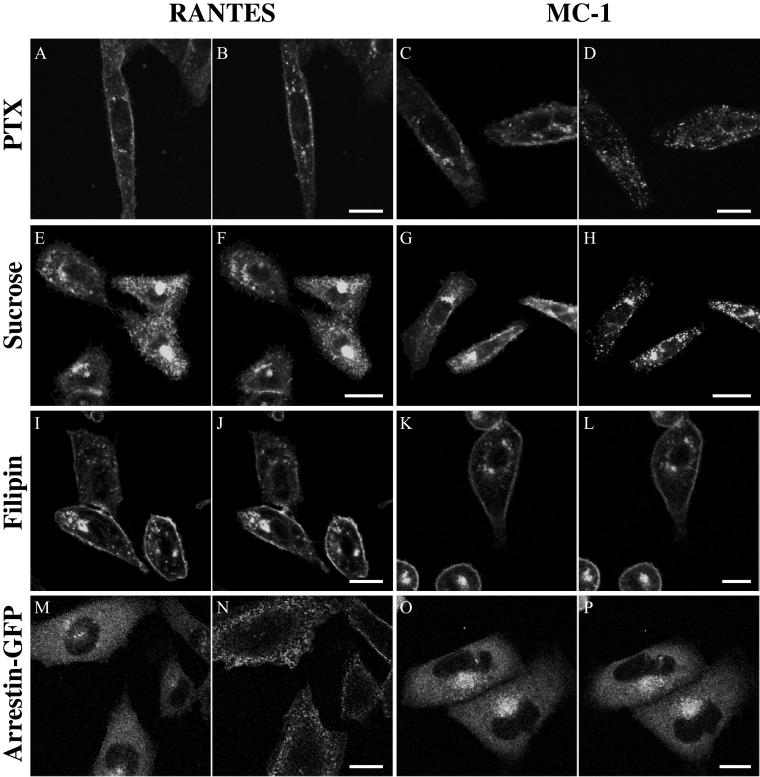
Figure 7.
MC-1 induces CCR5 endocytosis by an arrestin and clathrin-independent, caveolae-dependent pathway. (A–L). CHO-K1 cells expressing CCR5-GFP were incubated for 18 h with 100 ng/ml pertussis toxin 100 (A–D) or for 1 h with 0.45 M sucrose (E–H), or or 2.5 μg/ml filipin (I–L) for 1 h at 37°C. Cells were then stimulated with 100 nM RANTES (A, B, E, F, I, and J) or 10 μg/ml MC-1 (C, D, G, H, K, and L), and the dynamics of CCR5-GFP was recorded by confocal microscopy. Responses are shown before (A, C, E, G, I, and K) and 30 min after (B, D, F, H, J, and L) ligand addition. (M–P). CHO-K1 cells stably expressing CCR5 were transfected with β-arrestin-GFP; 24 h after transfection, subcellular redistribution of β-arrestin-GFP was analyzed by confocal microscopy. The cells were exposed to 100 nM RANTES (M and N) or 10 μg/ml MC-1 (O and P). Images were acquired every 15 s for 10 min. Responses are shown before (M and O), and 2 min (N) or 10 min (P) after ligand addition. Videos of the dynamics of β-arrestin-GFP trafficking in response to RANTES stimulation, as described in this figure, are available in the online version of this article. All experiments were repeated at least twice with similar results. Bar, 20 μm.

