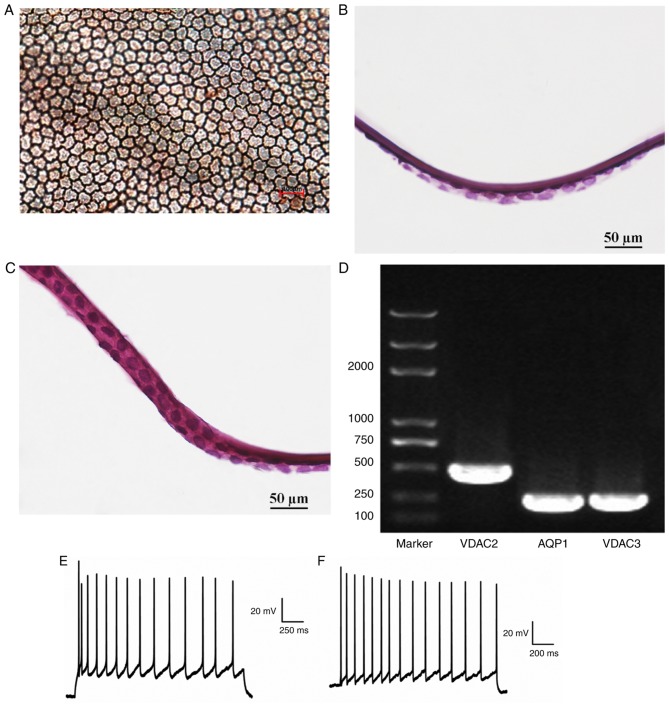
Figure 8.
Confirmation of successful RCEC implantation. (A) The implanted RCECs were stained with alizarin red-trypan blue. (B) Experimental group RCECs were seeded and tightly adhered onto the porcine DM endothelium surface. (C) Control group RCECs were tightly adhered onto the DM endothelium surface of fresh RCEC-DM complexes. (D) Electrophoresis results demonstrate expression of VDAC2, VDAC3 and AQP1 in implant RCECs. The action potential amplitudes of the (E) experimental and (F) control groups. RCEC, rabbit corneal endothelial cell; AQP1, aquaporin 1.

