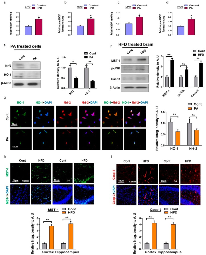
Figure 1.
Metabolic dysregulation induced oxidative stress both in in vivo and in vitro models: (a,b) The histogram showing the results of lipid peroxidation (LPO) and reactive oxygen species (ROS) levels in the high-fat diet (HFD) mice model; (c,d) a histogram represent the results of LPO and ROS levels in palmitic acid treated HT22 cells; (e) shown are the Western blot results of nuclear factor-2 erythroid-2 (Nrf-2) and hemeoxygenase-1 (HO-1) along with respective histograms in the palmitic acid-treated HT22 cells. β-Actin was used as a loading control; (f) shown are the Western blot results of mammalian sterile 20-like kinase-1 (MST1), phosphor-c-Jun N-terminal Kinase (p-JNK), and Caspase-3 along with respective histograms in brain homogenates of HFD-fed mice and the normal control group. β-Actin was used as a loading control; (g) representative images of immunofluorescence staining of colocalization of Nrf2/HO-1 in palmitic acid-treated cells; (h,i) immunofluorescence staining images of MST1 and Casp-3 in mice cortex and the hippocampus region. n = 12 mice/group. The data are shown here as a mean ± SEM. * p < 0.05, ** p < 0.01.

