
Figure 6.
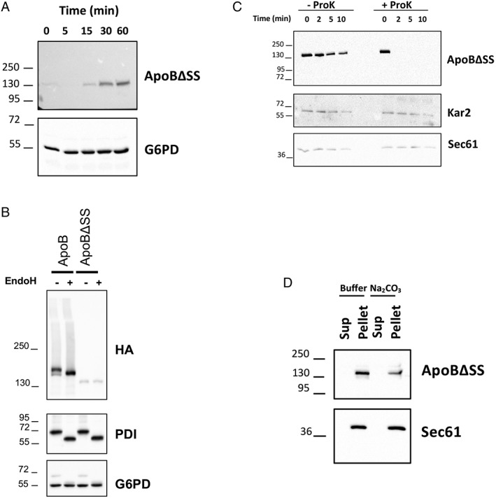
The signal sequence‐containing ApoB species translocates into the ER, whereas the signal sequence‐deficient protein remains tightly associated with the external surface of the ER. (A) Rapid expression of the signal sequence‐deficient protein (ApoBΔSS) after addition of β‐estradiol. An identical pattern of expression was observed when the signal sequence‐containing species was examined (Supporting Information Fig. S4A). G6PD served as a loading control. (B) Lysates from cells expressing ApoB and ApoBΔSS were treated with buffer or EndoH, as indicated, proteins were resolved by SDS‐PAGE, and ApoB (HA), the glycosylated PDI protein (as a positive control), and G6PD (as a negative control) were detected by Western blotting. (C) ER‐derived microsomes from cells expressing ApoBΔSS were treated with buffer or proteinase K (ProK), as indicated, and the stability of the proteins as well as the ER lumenal protein Kar2 and the integral ER membrane protein Sec61 (as negative controls) were examined by western blotting. (D) A crude membrane fraction from cells expressing ApoBΔSS was treated with buffer or sodium carbonate, and after centrifugation, the residence of the protein as well as Sec61 (as a positive control) in the supernatant (Sup) or pellet fraction was analyzed by Western blotting.