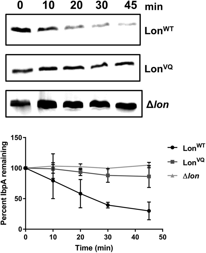
Figure 5.

In vivo degradation of IbpA by Lon upon heat shock. Representative Western blots of E. coli cell extracts during heat shock (45°C) monitoring loss of the IbpA protein over time. Bottom. Protein bands were quantified and the percentage of IbpA protein remaining when LonWT (circles), LonVQ (squares), or Δlon (triangles) was expressed is plotted as a function of time. The experiment was performed in triplicate and error bars represent SEM (lines connecting data points are not indicative of statistically based fitting).
