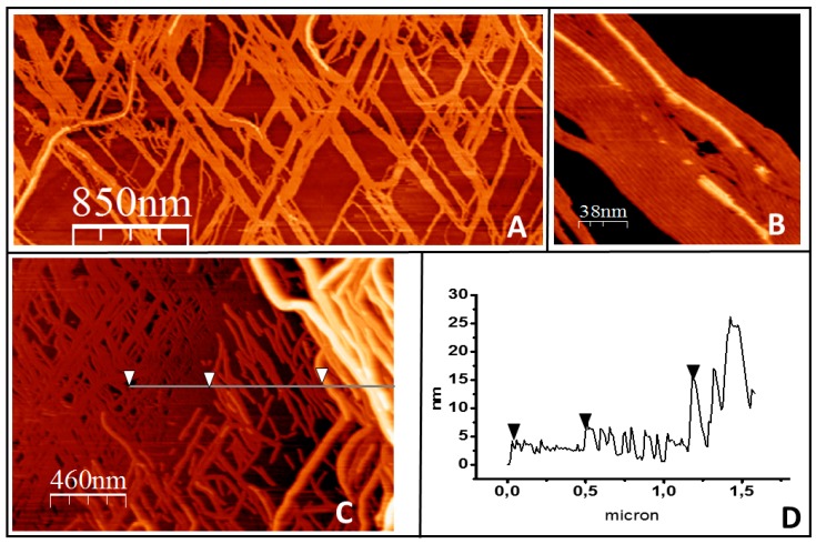
Figure 4.
Filament growth of tightly bound protein in three dimensions. (A) shows a general overview of filament bundles on the lipid surface. (B) shows how the filaments growing on top closely follow the orientation of the filaments below. (C) illustrates that in the areas where the lipids become fully covered by the protein, the second protein layer develops into ever higher and more complex structures. The height profile of the line is represented in (D). The arrows in both panels are a guide to show the three height levels observed: the first layer of filaments, the second layer, and a third layer of thicker structures.

