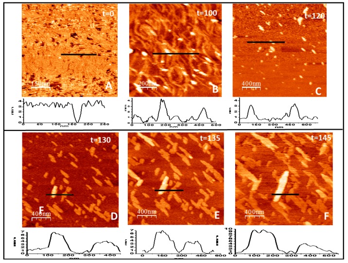
Figure 5.
Filament growth of loosely bound protein in two and three dimensions. (A–F) show the time evolution (in minutes) of the structures formed by FtsZ attached to the lipid surface through a flexible PEG linker immediately after GTP addition (t = 0). The graph below each image represents the topographical profile acquired at the position indicated by the horizontal line (all numbers are in nm). Well defined individual filaments formed immediately after GTP addition (A) condense into thicker curved bundles (B) that later disaggregate, leaving only some agglomerates that retain the height of 4 nm of the filaments (C). After a few minutes, condensation takes place again (D) and leads to the formation of linear arrangements that recover the height of one and two layers of lateral filament bundles that grow both longitudinally and laterally at a defined orientation (E,F).

