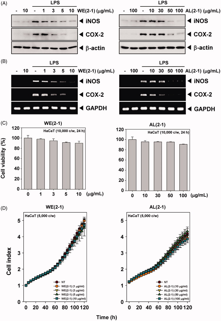
Figure 3.
Inhibition of the WE(2-1) and AL(2-1) on LPS-induced iNOS, and COX-2 gene products in RAW 264.7 macrophages. (A) RAW 264.7 cells were pre-treated with the indicated concentrations of WE(2-1) and AL(2-1) for 2 h before being incubated with LPS (1 μg/mL) for 22 h. Total RNA was isolated, and iNOS and COX-2 mRNA expressions were examined by RT-PCR analysis. PCR of glyceraldehydes-3-phosphatedehydrogenase, GAPDH, was performed to control for a similar initial cDNA content of the sample. The results shown are representative of the three independent experiments. (B) RAW 264.7 cells were pre-treated with different concentrations of WE(2-1) and AL(2-1) for 2 h and stimulated with LPS (1 µg/mL) for 22 h. Equal amounts of total proteins (10 μg/lane) were subjected to 10% SDS-PAGE, and the expressions of iNOS and COX-2 proteins were detected by Western blotting using specific anti-iNOS and anti-COX-2 antibodies. β-actin was used as a loading control. The blots shown are representative of three independent experiments that had similar results. (C) HaCaT cells (1 × 10⁴ cells/well) were treated with the indicated concentrations of WE(2-1) and AL(2-1) for 24 h and cell viability was determined by MTT assay. Results of independent experiments were averaged and are shown as percentage cell viability compared with the viability of untreated control cells. (D) Cell proliferation assay was performed using the Roche xCELLigence Real-Time Cell Analyzer (RTCA) DP instrument (Roche Diagnostics GmbH, Germany) as described in ‘Material and methods’. After HaCaT cells (5 × 10³ cells/well) were seeded onto 16-well E-plates and continuously monitored using impedance technology.

