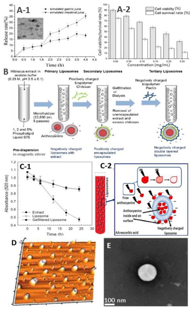
Figure 4.
Different panels showing in vitro release rate (A1) and absorption efficiency using the Caco-2 cell model (A2) with the inset in A1 showing TEM image of cyanidin-3-glucoside nanoliposome, schematic representation of multilayered anthocyanin nanoliposome prepared from hibiscus (B), the physical stability of black carrot anthocyanin nanoliposome and extract in the presence of ascorbic acid (C-1) along with the proposed mechanism of anthocyanin protection (C-2), and atomic force microscopy (AFM) (D) and TEM (E) images of anthocyanin nanoliposomes prepared from pistachio hull and bilberry extracts, respectively (adapted with permission from references [65,68,69,70,71]).

